Epilepsy treatment
The main aim of treating epilepsy is to improve the person's quality of life by preventing seizures but also causing minimum side effects
Getting a diagnosis of epilepsy can often take some time. Only until there have been two or more seizures that a diagnosis of epilepsy will be considered, unless there is an obvious known cause.
There is no test for epilepsy and diagnosis is almost entirely dependent on medical history, although can be possible with an accurate eye witness account.
It can be a long and frustrating process for families, so it is important to keep accurate records of any seizures, noting down when they occur, how long they last and a full description of what happened, including any changes in behaviour before the seizure. This will help the child’s medical team to understand the full picture.
Usually an initial medical examination will take place and the results of any further investigations will also take into account eyewitnesses reports.
NB: Young Epilepsy are currently reviewing our information portfolio, and will be updated October 2023.
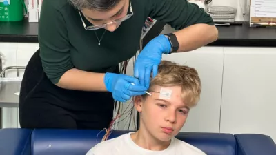
An EEG or electroencephalogram records the electrical activity in the brain.
It is a non invasive, painless procedure where electrodes, about the size of shirt buttons, are placed on the head and the signals are recorded by a computer. It usually takes place in a hospital or clinic.
The results of the EEG only give information about the electrical activity of the brain during the period of recording, it does not prove diagnosis of epilepsy.
Many people with epilepsy will have a normal trace. Only if specific patterns or characteristics of epilepsy are seen during the routine recording, is the EEG of value.
There are lots of different types of EEG testing:
A routine EEG can be useful for anyone with a diagnosis, or suspected diagnosis of epilepsy. A short recording (up to 45 minutes) is taken whilst the person is awake.
During a routine EEG activation procedures are often performed which are used to obtain more information which cannot always be seen on an EEG whilst just sitting quietly. These activation procedures can include; deep breathing, photic stimulation (where a flashing light is watched at different flash rates), exercise, sleep deprivation and drug reduction.
Once the electrodes are in place, the person may move around freely during an ambulatory EEG. The information that the electrodes is recording is stored on a small recorder and downloaded on to a computer at the end.
This type of EEG lets the person sleep in normal conditions, and allows day to day activities to continue. Sometimes a portable video camera can be used with the ambulatory EEG.
The recording may be performed for up to a week; however 24 to 48 hours are more usual.
Link to our health services
Video telemetry involves video monitoring taking place at the same time as the EEG recording.
The main advantage over the ambulatory EEG is that the person’s activities and movements are recorded at the same time as their brainwaves. Another advantage is that additional electrodes can be positioned elsewhere for a more detailed recording.
A disadvantage of video telemetry is that the person will need to spend most of their time in the same room, which can be quite tedious especially for young children.
Although this test can continue for up to a week, it usually lasts between 1-3 days. A parent or guardian must accompany the person having telemetry.
As part of our epilepsy assessment service, children and young people are given a diagnostic assessment in our video telemetry suite.
Some children will have brain scans which may help identify a cause of epilepsy.
This produces very detailed cross-sectioned images of the brain onto a computer.
CT scans show both bone as well as soft issues including the various areas of the brain. The scan may reveal any obvious structural abnormality or damage.
Using magnetic fields, rather than X-rays, this scan forms an image of the structure of the brain and is more detailed than a CT scan.
It is the most common scan type for children as it is sharper, and can reveal far smaller structural abnormalities. The disadvantage is that it takes longer and is noisy.
Some children may need sedation or anaesthesia prior to an MRI scan as they will have to lie still for some time. They may also be unsettled by the noise or claustrophobic feeling of being in the scanner.
An MRI scan with functional imaging (fMRI) is able to look at both the brain structures and areas of abnormality, as well as the blood flow in a specific area of the brain.
Actions such as thought, speech, movement and sensation cause an increase in the blood flow to the area in the brain that is activated. This makes the fMRI extremely useful for mapping the brain for its various functions and for locating where seizures start.
During the test a computer collates the images of the brain and shows them in cross sections. These can be added together to form a 3D image.
Because a SPECT scan can show areas of reduced blood flow, it is more likely to pick up brain injury because there is likely to be reduced blood flow to the affected area.
During this test the child is given injection into the blood stream. When scanning is then performed, the blood flow through the arteries and veins in the brain can be traced. If this is done during a seizure, it will show where in the brain the seizures started.
PET scans are more precise than SPECT scans. They are able to show how tissues in the brain are functioning and show areas which may be structurally abnormal, that do not show up on an MRI scan. These scans are not widely available.
Various blood tests may be needed either to exclude other diagnoses or in an attempt to find the underlying cause of the epilepsy.
If these tests are needed, the blood required for the tests can usually be taken in one go
Diagnosing epilepsy can be difficult, as there are a wide range of conditions that look like epileptic seizures.
Sometimes faints (syncopal attacks) can have jerking movements and are often mistaken for seizures. Other conditions that can look similar to epilepsy include:
This condition is not a type of epilepsy but can occur in people who have epilepsy.
Someone with NEAD has attacks that look similar to epileptic seizures, but they are not caused by abnormal electrical activity in the brain or by another medical cause. NEAD attacks can cause black outs, falls, loss of bladder control and are associated with injuries.
If a child has a suspected seizure, it is likely that they will be taken to the nearest Accident and Emergency department or in some cases they will be seen by their GP.
It may be that the doctor thinks that the child may have had an epileptic seizure, in which case, an urgent appointment with a specialist (a paediatrician who has had special training in diagnosing and treating epilepsy) should be made within two weeks.
The paediatrician will refer on to a specialist centre if there is uncertainty about the diagnosis, where the child will be seen by a paediatric neurologist.
The main aim of treating epilepsy is to improve the person's quality of life by preventing seizures but also causing minimum side effects
An epileptic seizure happens because of a disruption of the electrical activity in the brain
Knowing what can cause a seizure can help to manage epilepsy
A syndrome is a group of signs and symptoms which, if they occur together, can suggest a particular condition
Sudden unexpected death in epilepsy (SUDEP) is extremely rare and affects only around 500 people in Britain every year
There are certain conditions or disorders that may, or may not, accompany epilepsy. These are sometimes known as co-morbidities
It is important that all teenagers are given information about the effects of AEDs on contraception and pregnancy before they become sexually active
The causes of epilepsy generally falls into three groups; genetic, structural/metabolic or unknown